INFORMATION ABOUT HUMAN BODY.
Monday, 26 August 2013
COMBINATION OF MECHANICAL & ELECTRICAL: A.C. Induction Meter
COMBINATION OF MECHANICAL & ELECTRICAL: A.C. Induction Meter: . A. C. induction-type energy meters are the most common form of meters met within everyday domestic and industrial installations. T...
Tuesday, 21 May 2013
THE CORNEA.
A structure at the outer angle of the antericr chamber, the intervals between the fibres forming small cavernous spaces, the spaces of Fontana. 'fhese little recesses communicate with a circular canal in the substance of the s(llerotic
close to its junction with the cornea. This is the canal of Schlemm, 01' sinus t'en08US 8cler(£; it communicates internally with the anterior chamber through the spaces of Fontana,and externally with the scleral veins. Some of the fibres of this reticulated structure are continued into the front of the iris, forming the ligamentum pectinatum iridi8; while others are connected with the fore part of the sclerotic
. The nerves are numerous, twenty-four to thirty.six in number (Kolliker), forty to forty.five (Waldeyer and Sumisch); they are derived J from the ciliary nerves and enter the laminated tissue of the cornea. They ramify thrQughout its substance in a delicate network, and their terminal fila¬ments fQrm a firm and closer plexus on the surface of the cornea proper beneath the epithelium.
THE BRAIN
According to him, the weight of the brain has no in~ue.nce whatever on. the mental faculties. It ought to b& remembered that the slgmficance of the weight of the brain should depend upon the proportion it bears to the dimensions of the whole body and to the age of the individual. It is equally important to know what was the cause of death, for long illness or old age exhausts the brain. To define the real degree of development of the brain it is therefore necessary to have a knowledge of the condition of the whole body, and, as this is usually lacking, the mere record of weight possesses little significance.
The human brain is heavier than that of all the lower animals, excepting the elephant and whale. The brain of the former weighs from eight to ten pounds; and that of a whale, in a specimen seventy-five feet long, weighed rather more than five pounds.
Cerebral Localization and Topography.-Physiological and pathological research have now gone far to prove that the surface of the brain may be mapped out into series of definite areas, each one of which is intimatelv connected with some well-defined function. And this is especially true with reeard to the convolutions on either side of the fissure of Rolando, which are believed by most pbysiolozists of the present day to be concerned in motion, those grouped around the fissure being associated with movements of the extremities of the opposite side of the body, and those around the lower end of the fissure being related to movements of the mouth and tongue
THE TONGUE.
The tongue, being very vaRcular, is often the seat of neevoid growths,and these have a tend-ency to grow rapidly.The tongue is frequently the seat of ulceration, which mav arise from many causes, as from
the irritation of jagged teeth, dyspepsia, tubercle, syphilis, and cancer. Of these the cancerous ulcer is the most important, and probably also the most common. The variety is the squamous epithelioma, which soon develops into an ulcer with an indurated base. It produces great pain, which speedily extends to all parts supplied with sensation by the fifth nerve, especiall;v to the region of the ear. The pain in these cases is conducted to the ear and temporal region by the lingual nerve, and from lt to the other branches of the inferior maxillary nerve, especially the auriculo-temporal. Possibly pain in the ear itself may be due to implication of the fibres of the glosso-pharyngeal nerve, which by its tympanic branch is conducted to the tympanic plexus. .
Cancer of the tongue may necessitate removal of a part or the whole of the organ, and many different methods have been adopted for its excision. It may be rerum'ed from the mouth by the ecraseur or the scissors. Probably the better method is by the scissors. usually known as Whitehead's method. The mouth is widely opened with a gu,!!. the toncue transfixed with a stout silk ligature, by which to hold and make traction on it and the reflection of mucous membrane from the tongue to the jaw, and the insertion of the Genio-hyo-glossus first divided with a pair of curved blunt scissors. The Palato-glossus is also divided. The tongue can now be pulled well out of the mouth. The base of the tonzue is cut through by a series of short, snips, each bleeding vessel being dealt with as soon as divided, until the situation of the ranine arterv is reached. The remaining undivided portion of tissue is to be seized with a pair of 'Yells's forceps, the tongue removed, and the vessel secured. In the event of the ranine artery being accidentally injured hremorrhage can be at once controlled by passing two fingers over the dorsum of the tongue as far as the epiglottis and dragging the root of the tongue forcibly
forward.
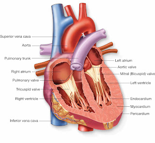
THE HEART
Component Parts.-As has already been stated, the heart is sub¬divided by a muscular septum into two lateral halves, which are named respectively right and left; and a transverse constriction subdivides each half of the organ into two cavities, the upper cavity on each side being called the auricle, the lower the ventricle. The course of the blood through the heart cavities and blood-vessels has already been described
The division of the heart into four cavities is indicated by grooves upon its surface, The groove separating the auricles from the ventricles is called the auriculo-ventricular groove. It is deficient, in front, where it is crossed by the root of the pulmonary artery. It contains the trunks of the nutrient vessels of the heart. The auricular portion occupies the base of the heart, and is su bdivided into two cavities by a median septum. The two ventricles are also separated into a right and left by two furrows, the interventricular grooves, which are situated one on the anterior, the other on the posterior, surface; these extend from the base of the ventricular portion to near the apex of the organ; the former being situated
Thursday, 16 May 2013
EYE
This is the colored part of the eye: brown, green, blue, etc. It is a ring of muscle fibers located behind the cornea and in front of the lens. It contracts and expands, opening and closing the pupil, in response to the brightness of surrounding light. Just as the aperture in a camera protects the film from over exposure, the iris of the eye helps protect the sensitive retina.
These two Iaminee are connected by an intermediate stratum; which is destitute of pigment-cells and consists of fine elastic fibres. On the inner surface of the lamina chorio-capillaris is a very thin, structureless, or, according to Kolliker, faintly fibrous membrane, called the lamina basalis or membrane of Bruch; it is closely connected with the stroma of the choroid, and separates it from the pigmentary laver of the retina .
•Tapetum.-This name is applied to the iridescent appearance which is seen in the outer and posterior part of the choroid of many animals.
The ciliary body should now be examined. It may be exposed, either by detaching the iris from its connection with the Ciliary muscle, or by making a transverse section of the globe, and examining it from behind.
The ciliary body comprises the orbiculus ciliaris, theciliary processes, and the Ciliary muscle.
The orbiculus ciliaris is a zone of about one-sixth ofan inch in width, directly continuous with the anterior part of the choroid; it presents numerous ridges arranged in a radial manner.
The ciliary processes are formed by the plaiting and folding inward of the various layers of the choroid-i. e., the choroid proper and the lamina basalis-at its anterior margin, and are received between corresponding foldings of the suspensory ligament of the lens, thus establishing a connection between the choroid and inner tunic of the eye. They are arranged in a circle, and form a sort of plaited frill behind the iris, round the margin of the lens. They vary between sixty and eighty in number, lie side by side, and may be divided into large and small j , the latter, consisting of about one-third of the .entire number, are situated in the spaces
THE ORGANS OF DIGESTION
The muscles of the soft palate are five on each side: the Levator palati, Tensor palati, Azygosuvulm, Palato-glossus, and Palato-pharyngeus . The following is the relative position of these structures in a dissection of the soft palate from the posterior or nasal to the anterior 01' oral surface: Immediately beneath the nasal mucous membrane is a thin stratum of muscular fibres, the posterior fasciculus of the Palato-pharyngeus muscle, joining with its fellow of the opposite side in the middle line. Beneath this is the Azygos uvulm, consisting of two rounded fleshy fasciculi, placed side by side in the median line of the soft palate. Next comes the aponeurosis of the Levator palati, joining with the muscle of the opposite side in the middle line. Fourthly, the anterior fasciculus of the Palato-pharyngells, thicker than the posterior, and separating the Levator palati from the next muscle, the Tensor palati. This muscle terminates in a tendon which, after winding round the hamular process, expands into a broad aponeurosis in the soft palate, anterior to the other muscles which have been enumerated. Finally, we have a thin muscular stratum, the Palato-glossus muscle, placed in front of the aponeurosis of the Tensor palati, and separated from the oral mucous membrane by adenoid tissue.
The Tonsils (amygdalce) are two prominent bodies situated one on each side of the fauces, between the anterior and posterior pillars of the soft palate. They are of a rounded form, and vary considerably in size in different individuals. A recess, the fossa supra-tonsillaris, may be seen, directed upward and backward above the tonsil. His regards this as the remains of the lower part of the second visceral cleft. It is covered by a fold of mucous membrane termed theplica triangularis. Externally the tonsil is in relation with the inner surface of the Superior constrictor, to the outer side of which is the Internal pterygoid muscle. The internal carotid artery lies behind and to the outer side of the tonsil, and nearly an inch distant from it. It corresponds to the angle of the lower jaw. Its inner surface presents from twelve to fifteen orifices, leading into small recesses, from which numerous follicles branch out into the substance of the gland. These follicles are lined by a continuation of the mucous membrane of the pharynx, covered with epithelium; around each follicle is a layer of closed capsules imbedded in the submucous tissue. These capsules are analogous to those of Peyer's glands, consisting of adenoid tissue. No openings from the capsules into the follicles can be recognized. They contain a thick grayish secretion. Surrounding each follicle is a close plexus of lymphatic vessels. From these plexuses the lymphatic vessels pass to the deep cervical giands in the upper part of the neck, which frequently become enlarged in affections of these organs.
Subscribe to:
Comments (Atom)


